Inextricably tied to orthopaedics are the basic sciences. This fundamental concept has governed product development at OSI from 1999 through today. We approach each innovation with a sense of realism and not optimism. Diabetes, hypertension, and obesity are common in orthopaedics, as are declining reimbursements. Notably, when things do not go well, there is always a shortage of persons responsible. Orthopaedists need innovative products that minimize risks, not more products that mitigate risks, as for each level of mitigation, there are more costs and more risks. The status quo is expensive, layered, and has become a bulwark to the physician’s intellectual relevance. OSI recognizes that in orthopaedics, how great a product is matters less if the treating physician cannot perceive the benefits intended for the patient. The information provided herein is informative and aims to help one think more broadly about OA and AVN by offering products that are “problem up solutions,” meaning products that provide solutions to the specific clinical challenges one might face. A great practice is different from a great outcome, and innovative medical devices are needed to address this binary through the alignment of the incentives of all stakeholders.
Our products are NOT a substitute for an arthroplasty; they are products for patients seeking sustainable joint preservation solutions during the run-up to an arthroplasty and are based on the orthopaedic basic sciences. While each of our products may be used as a stand-alone device, OSI encourages the physician to think more broadly about the etiology of osteoarthritis and question the perpetual commentary of “osteoarthritis of unknown etiology.” That said, when one has a deeper understanding of OA and AVN, the usefulness of our products becomes more apparent.
Carefully review the cited literature.1-22 Consider the significant and well-known role of oxidized low-density lipoproteins (oxLDL) in cardiovascular disease. Importantly, these data are published in journals not commonly read by orthopaedists. In CORR in 1948, Fremont Chandler captured this then-unknown relationship when describing AVN as a coronary of the hip. Then in 1985, Brown and Goldstein received the Nobel Prize for characterizing cholesterol metabolism. Since their seminal work, much has been learned about plaque formation in the coronary arteries. More importantly, recent findings have shown the presence of intraosseous oxLDL in arthritic joints. In 2002, Brannon did NOT assign his endoscopic findings of calcified marrow spaces within the femoral head as an artifact, nor was he persuaded by those who did and perhaps still do today. He questioned conventional wisdom, sought answers, and repeatedly confirmed AVN on histopathology. Brannon used his solid knowledge of biochemistry, physics, cell biology, and the orthopaedic basic sciences to understand and explain these new observations at the cellular level. The answers obtained allowed OSI to develop products that facilitate the restoration of articular congruency, the release of accumulated intraosseous fat, oxLDL, associated proinflammatory cytokines, and the debridement of necrotic bone within the femoral head. Thus, restoring the biology of the bone and joint before its form and function are lost. With this more profound understanding of OA and AVN, it is apparent that an intraosseous injection of a spun down bone marrow aspirate, PRP, if ever contemplated, or dual therapy bisphosphonates could not embrace the published literature and only support the use of “existing” products in a so-called novel way. Further, viscosupplements have been repeatedly shown to be ineffective.
While any reader hereof can immediately contemplate the success of a given technique in their practice or that of a surgeon more broadly known, the etiology of OA remains unknown, and its shared pathophysiology with AVN has not been considered. This is the fundamental issue that drives product development at OSI. Intraosseous fat and oxLDL-induced release of cytokines and chemokines play a major role in disease. While significant intellectual energy has been devoted to understanding the mechanics of a replaced knee at the bone prosthesis interface, one cannot ignore the reasoning that offending cytokines, chemokines, and oxLDL are released during the preparation of the knee for its replacement. Further, nothing prevents reaccumulation of these biomarkers after the knee is replaced and their reaccumulation may lead to aseptic loosening. Frankly, we have not considered the value of a total knee in this physiologic direction. That said, a major joint can be viewed as the end-organ of a long bone, as did Fremont Chandler. Thus, once metabolic products or by-products are deposited at the ends of long bones, they are not easily removed. A willingness to review the cited literature and think more broadly in this regard allows one to consider how a patient may experience an oxLDL storm upon release of the tourniquet during a total knee, and how high levels of LDL combined with increased levels of reactive oxygen species due to the oxidative stress of surgery may pose a substantial risk for a postoperative DVT.
Review our products in detail, ask questions, request more information, and request a video demonstration. Be a true skeptic, question everything we have presented on our web page as information, and consider adding oxLDL levels to the panel for your joint fluid analyses in patients you see with OA and an effusion in your office. Perform a core decompression and send the core sample to pathology and visit with your pathologist. Endoscopically examine the core track to observe the calcified marrow spaces before and after a degree of debridement, and you decide if the findings are artifacts. Does subchondral sclerosis comprise thickened trabeculae or trabeculae with calcified marrow spaces? Review David Helfet’s cadaver study of the femoral head wherein he and others describe its transosseous blood flow and how the foveal, superior retinacular and inferior vincula arteries communicate transosseously through a capillary network. Look inside of the femoral head if you have not done so previously. The inability to observe calcified marrow spaces and completely map their location in a case of adjudicated AVN results from not using the proper equipment and the clinical axiom of the AVN lesion is circumscribed by a sclerotic margin, according to Steinberg. Thus, most core decompression systems ream away the bone antecedent to the lesion, losing an opportunity to better understand the disease. Heretofore, no one has ever been able to endoscopically observe the necrotic bone circumscribed by a sclerotic margin and compare it to the bone immediately inferior to the sclerotic margin. This simple comparison reveals that the necrotic bone within the sclerotic margin is nearly histopathologically identical to the bone immediately inferior to the sclerotic margin, i.e., both areas demonstrate bone with occluded marrow spaces. This raises the question of what causes the marrow spaces to calcify and what articular changes occur inside of the joint when the transosseous blood flow is obstructed? Review Freund's 1936 paper on avascular necrosis. Make time to visit your lab, look at the histology of the tissue specimens you obtain when treating AVN, and compare these observations to the preoperative MRI. Is there concordance? Ask more questions. Does oxLDL induce calcification of the marrow spaces in a manner similar to calcification of foam cells in cardiovascular disease? Read the references below. Does oxLDL destroy cartilage and induce inflammation? How does oxLDL gain access to the joint? In the case of the knee, does oxLDL enter the joint through rupturing osteophytes? Is there a hereditary component relative to the quantity and anatomic distribution of oxLDL receptors, LOX-1 (lectin-like oxLDL receptor 1), found on endothelial cells and cartilage in a given joint? LDL has a high affinity for the proteoglycan biglycan, which is encoded on the X chromosome. Does the gender distribution of biglycan help explain why OA is more common in a female? What role do SR-1 oxLDL receptors on osteoclast play in bone resorption? Notably, it is well-admitted in the peer-reviewed published literature that oxLDL receptors are located on platelets and macrophages. When performing a total knee arthroplasty and instrumenting the medullary canal, retain a fat sample to analyze it for oxLDL. In the case of early aseptic loosening after a total knee, cemented or press-fit, could the reaccumulation of fat and oxLDL, having strong pro-inflammatory properties, lead to early aseptic loosening of the prosthesis? Autophagy plays a major role in aseptic loosening, but when described by the orthopaedic scientific community, no consideration is given to the role oxLDL may play in autophagy. How could wear particles account for early loosening? In cases of aseptic loosening, sample the periprosthetic joint fluid and the intraosseous bone space and analyze it for oxLDL. Measure systemic and local oxLDL levels at the time of an aseptic revision. Yes, be concerned about the return of blood flow during a total knee arthroplasty when the tourniquet is deflated in a patient with peripheral vascular disease, but ask questions regarding the similarities between calcified marrow spaces and calcified arteries. Review the mineralization mimicries between calcifying vascular smooth muscle cells and osteoblasts. Ask questions about the similarities between oxLDL-induced plaque formation in the intima of the popliteal artery and induced calcification within the marrow spaces of the arthritic knee. Do calcifications within the marrow spaces contribute to the formation of sclerotic subchondral bone? Is there a tremendous systemic load of oxLDL (oxLDL Storm) released into the bloodstream when the tourniquet is deflated? Fat is dispersed intraosseously when press-fitting a femoral stem, but what amount of this fat is oxLDL, and is it released into the circulation? Unoxidized LDL is not proinflammatory. However, oxLDL is proinflammatory along the endothelial surface, which is well-admitted in the cardiac literature. Does circulating oxLDL released from the bone cause inflammation of the endothelial lining and lead to postoperative DVTs and MIs? Can preoperative and postoperative oxLDL levels serve as a biomarker and provide insight into those patients likely to develop a postoperative DVT or an MI? During fat embolization syndrome, what role does oxLDL play in inflammation of the pulmonary vasculature? When aspirating a prosthetic joint that is particularly inflamed, could the proinflammatory properties of oxLDL explain the untoward inflammation in the presence of negative cultures? In the case of aseptic loosening, could the percutaneous intraosseous irrigation of the periprosthetic bone remove oxLDL, proinflammatory cytokines, and macrophages with ingested wear debris to a degree such that new bone formation can occur when the periprosthetic area is infused with an autologous cancellous bone graft slurry and bone marrow? Review OSI’s Biomass Delivery Curette for this application. Notably, enthusiasm about a minimally invasive treatment option for a devastating problem should NOT lead to periprosthetic concentrated bone marrow injections nor PRP injections because neither removes the oxLDL and proinflammatory cytokines. Here again, such an application misses the fundamentals of intraosseous oxLDL and its proinflammatory properties. Is there an imbalance between RANKL-RANK and osteoprotegerin wherein adaptive new bone formation is compromised? Does oxLDL augment an already overloaded macrophage-induced inflammatory process in late-stage aseptic loosening? The answers to these questions are revealing and open vast opportunities for research and new product development. Could we mitigate against postoperative DVTs after a total knee by minimizing the amount of oxLDL released into circulation? OSI’s products don’t have all the answers, but we have the confidence to question conventional wisdom, and we are motivated by science, as our name implies. The success of an arthroplasty is not insignificant, but it is systemically irrelevant because the etiology of OA remains unknown.
We have not sought the views of a KOL to describe the benefits of our products. We encourage the orthopaedist to review the peer-reviewed published literature and then decide if our products can provide any value to their patients seeking joint preservation. Nothing in this reading can be construed to imply that the standard of care must change, but if a surgeon reader hereof is not motivated to ask questions about the role oxLDL may play in OA, AVN, Atypical AVN, its effects on osteoclastogenesis, and autophagy in aseptic loosening, nothing will ever be gained. For orthopaedic healthcare to improve, amongst its many other challenges, providers from a diverse workforce must share their knowledge and experiences to expand our collective understanding of osteoarthritis and AVN. A current query within the American Journal of Bone and Joint Surgery for “oxidized low-density proteins” and “oxLDL” produces zero results (see image below).
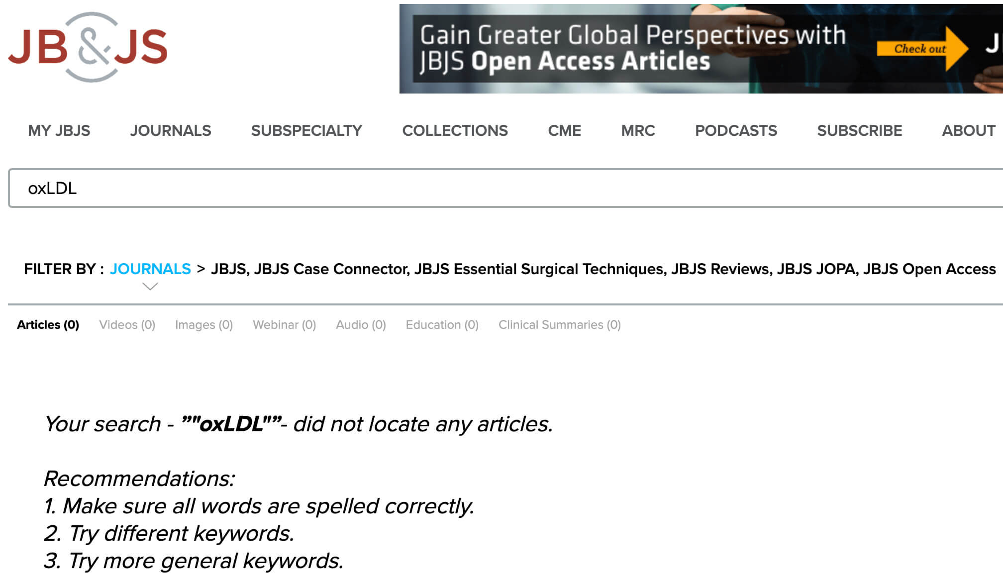
Figure 1: Recent search of JBJS reveals no published work on the oxidation of low-density lipoproteins. This implies that the proinflammatory role oxLDL may play through M1 macrophage release of IL-6, IL-8, CCL2, osteopenia, and insufficiency fractures has not been considered. Nonetheless, many consider these cytokines and CCL2 causative in autophagy-induced aseptic loosening through macrophage ingestion of wear debris. However, oxLDL is resident to the periprosthetic bone and reflects one's intake of saturated fats and trans fats and oxidative stress. That said, aseptic loosening may be due to synergism between the consequences of oxidative stress and the compounding effects of ingestion of wear debris. The proinflammatory properties of oxLDL are not restricted to the heart, and such metabolic activity does NOT stop when the joint is replaced.
Has something been missed? That said, the alternative is the status quo, which invariably deteriorates over time as costs soar, reimbursements decline, and regulations and burnout increase.
What one ultimately provides as joint preservation treatment for a patient is a clinical decision within one’s practice and may not include any of OSI’s products. However, we are confident that we have opened a new door to a broader understanding of arthritic disease, thus expanding treatment options for all.
This paper concludes that arthroscopy for osteoarthritis is ineffective. However, these authors describe inadequate debridement of the knee, in our view. Further, no description of meniscus function nor the release of the excess fat from the bone is described after debridement. The meniscus appears to have been removed in some cases. The paper describes the procedure as below:
“After diagnostic arthroscopy in patients in the débridement group, the joint was lavaged with at least 10 liters of fluid, rough articular cartilage was shaved (chondroplasty was performed), loose debris was removed, all torn or degenerated meniscal fragments were trimmed, and the remaining meniscus was smoothed to a firm and stable rim. No abrasion arthroplasty or microfracture was performed. Typically, bone spurs were not removed, but any spurs from the tibial spine area that blocked full extension were shaved smooth.”
This paper is a follow-up to the Moseley, et al. paper after the orthopaedic community considered its statistical methods inadequate. Nonetheless, Kirkley, et al. concluded that arthroscopy for osteoarthritis was ineffective after performing, what appears to be, inadequate debridement by using antiquated equipment, as in the Moseley paper. The paper describes the procedure as below:
“Arthroscopic treatment was performed within 6 weeks after randomization with the patient under general anesthesia and with the use of a tourniquet and a thigh holder. The orthopedic surgeon evaluated the medial, lateral, and patellofemoral joint compartments, graded articular lesions according to the Outerbridge classification, irrigated the compartment with at least 1 liter of saline, and performed one or more of the following treatments: synovectomy; débridement; or excision of degenerative tears of the menisci, fragments of articular cartilage, or chondral flaps and osteophytes that prevented full extension. Abrasion or microfracture of chondral defects was not performed.”
Mazière C, Louvet L, Gomila C, Kamel S, Massy Z, Mazière JC. Oxidized low-density lipoprotein decreases Rankl-induced differentiation of osteoclasts by inhibition of Rankl signaling. J Cell Physiol. 2009 Dec;221(3):572-8. DOI: 10.1002/jcp.21886. PMID: 19725047.
Camuzard, O; Breuil, V; Carle, GF.; Pierrefite-Carle, V. Autophagy Involvement in Aseptic Loosening of Arthroplasty Components. J Bone Joint Surg Am, 101(5):466-472 | Current Concepts Review | March 06, 2019.
Hashimoto K, Akagi M. The role of oxidation of low-density lipids in pathogenesis of osteoarthritis: A narrative review. J Int Med Res. 2020 Jun;48(6):300060520931609. DOI: 10.1177/0300060520931609. PMID: 32552129; PMCID: PMC7303502.